Back to sepiabio.com
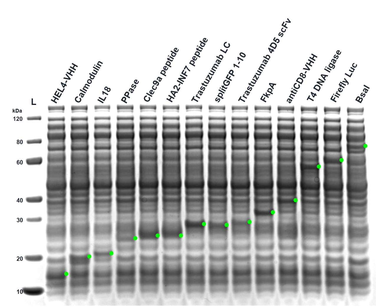
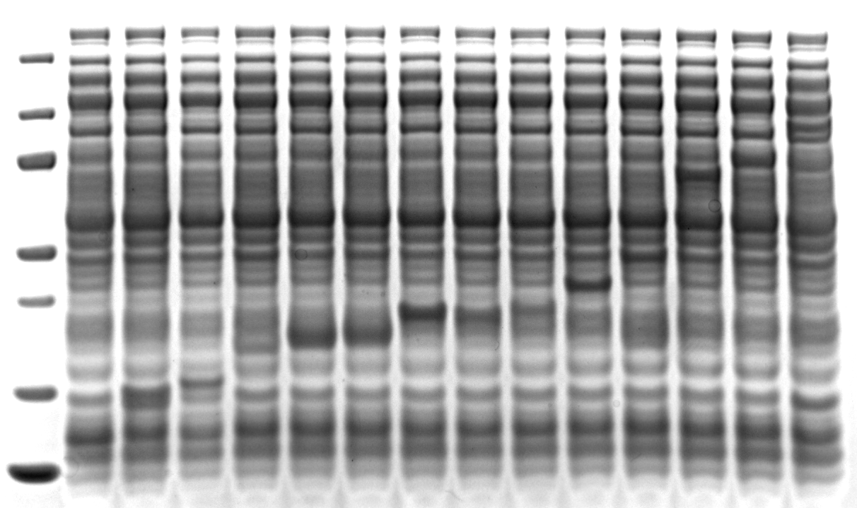
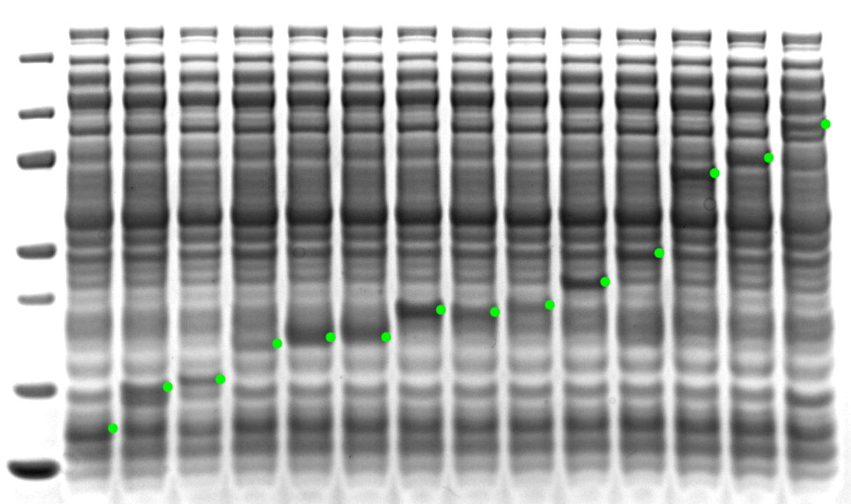
Figure 5. The OpenCFPS™ E. coli Core Kit produces a variety of proteins. Enzymes, antibody-derived biologics, peptide fusions, and other proteins of prokaryotic/phage, eukaryotic, or synthetic origin are expressed and visualized by running the soluble fraction (post-spin) of the resulting reaction on a Coomassie-stained SDS-PAGE gel.
For each construct, linear DNA cassettes were prepared that contain a ~100 bp spacer on both ends followed by a T7 expression promoter, strong ribosome binding site, the protein of interest, a terminator, and a Malachite green binding site. DNA was synthesized as fragments by Abstract Bio (California, USA) and/or Twist Biosciences (California, USA) and assembled by Golden Gate Assembly with enzymes and buffers from NEB (Massachusetts, USA) and an I.DOT DW (Dispendix, Stuttgart, Germany) followed by PCR amplification and silica-based Monarch Spin PCR & DNA Cleanup Kit (5 µg) (NEB). DNA size and purity was determined by agarose gel electrophoresis followed by A260 using a DS-11 FX+ (Denovix, Delaware, USA).
Cell-free reactions contained 30% Sepia Extract E0002 lot E000225121803, 40% Sepia Buffer B0001 lot B000126022403, linear DNA at 15-16 nM, 10 µM Malachite green (from a 1 mM stock), and water. Reactions were set up by DNA dispensation by an I.DOT DW and Master Mix and water dispensation by a Formulatrix Mantis v3.3 ACC RFID (Formulatrix, Massachusetts, USA). After dispensing, the plate was spun down, covered with a clear nonpermeable MicroAmp Clear Adhesive Film (ThermoFisher, California, USA) with a cut out for the wells to be read, then covered with a BreatheEasier film (Diversified Biotech, Massachusetts, USA). Reactions were conducted across 2 384-well plates at 10 µL scale, with one plate run in a Thermomixer C with flat plate adapter and cover (Eppendorf, Hamburg, Germany) to shake at 1200 rpm at 21°C and one plate in a Biotek Synergy Neo2 (Agilent, California, USA) with continuous orbital shaking (fast, 1 mm) at 21-24°C. The plate in the Neo2 was tracked at 610 ex / 650 em gain 140 (monochromator) in a kinetic read, with continuous orbital shaking (fast, 1 mm). Reactions were terminated at 12 h, combined together, and centrifuged at 21,130 × g for 5 min in an Eppendorf 5424 (Eppendorf, Hamburg, Germany).
The supernatant for a larger set of proteins was initially tested, the samples were frozen, and then after down-selection to a smaller set of proteins the samples were thawed. 1 µL of the supernatant was then run on a reducing 4-12% SurePAGE Bis-Tris Gel. Samples were denatured using LDS Reducing Agent and 50 mM DTT at 70°C for 10 min, and run with 5 µL of PAGE-MASTER Protein Standard (Genscript, Nanjing, China). The resulting gel was stained using Coomassie SafeStain (Invitrogen, California, USA) and imaged.
The following proteins are not shown after down-selection: KLF1, ProteinG-Tn5 (low soluble expression by FluoroTect (Promega, Wisconsin, USA)), Pfu polymerase (good soluble expression by FluoroTect but hidden by Coomassie stain), gamS, SUMO Protease, Snaptag-HA2long, Snaptag-HA2short, Snaptag-TatHA2, DsbC (21-236 aa), Snaptag2-meGFP (soluble expression by Coomassie stain but not selected due to space), and Gaussia luciferase, trastuzumab_Fab_HC, trastuzumab_HC (no soluble expression by Coomassie stain).
Protein structures were predicted locally using ColabFold/AlphaFold2 on a Mac mini with Apple M4 and 16 GB memory (Apple, California, USA), and secondary structures were determined by DSSP.
Click a protein to show its approximate expected-MW position on the SDS-PAGE gel.